
Indication
Most biliary imaging is non invasive with US, MRCP or CT.
Most percutaneous cholangiography is performed in the angio suite prior to intervention.
There are a few situations where you will need to perform cholangiograms.
To check patency of biliary drains (PTC)
To check patency of and leak from cholecystostomy tubes with imaging of the CBD
To check patency of the CBD following operative T-tube insertion
For all examinations you must answer the following questions before performing the procedure:
What is the clinical question to be answered?
What study will you perform?
How do you perform this study and what equipment is required?
Who will supervise or review your images when you need assistance?
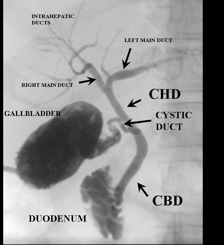
Anatomy
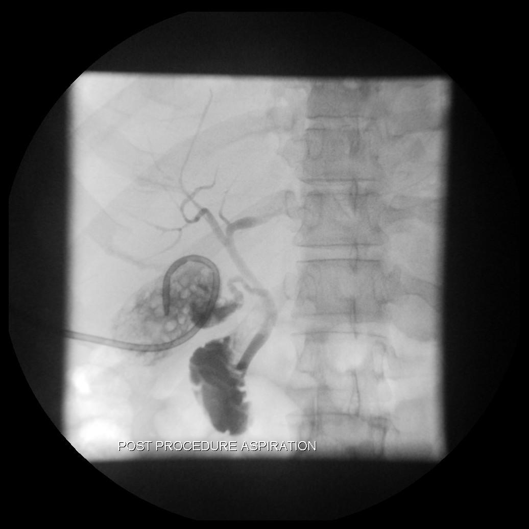
A percutaneous cholangiogram taken through a cholecystostomy tube
The pancreatic duct is rarely visualised during percutaneous cholangiography.
Equipment
Sterile pack, chlorhexadine, sterile towel, 20ml syringe x 2, saline, contrast (niopam).
Technique
This is a sterile procedure
As the drainage tube has often been inserted due to infection, most patients should be given antibiotics prior to imaging, or ensure sepsis has settled prior to imaging.
Clean and drape the appropriate tube.
Take a control film of the tube if visible. Often the distal end of the tube may not initially be visible. The nature of the tube, pathology and indication for cholangiogram will guide your control film.
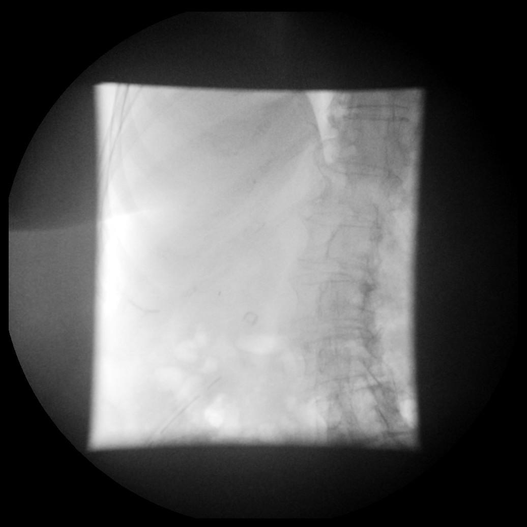
Control film – T Tube not visible
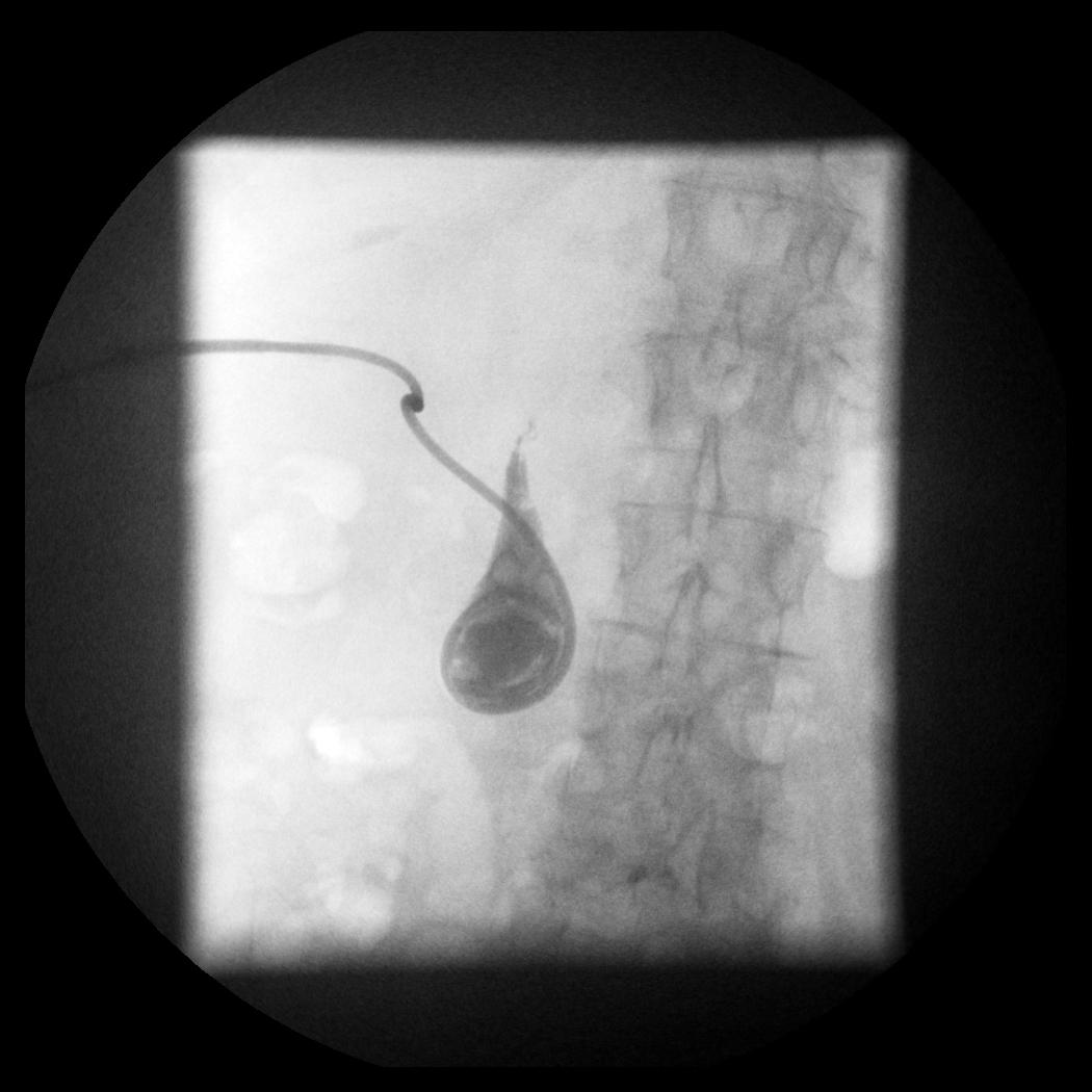
Gently aspirate some bile and flush a small volume of saline 3-5 ml to ensure the tube is patent. Ensure that you do not introduce air. Air bubbles will mimic calculi.
Gently inject Niopam using a small 10ml or 20 ml syringe. Start by opacifying just the tube using fluoroscopic guidance. You can make adjustments to your radiography i.e. centring, magnification and conning to optimise imaging. Take an exposure of the tube.
Early view showing filling of the T Tube. Now is the time to make adjustments.
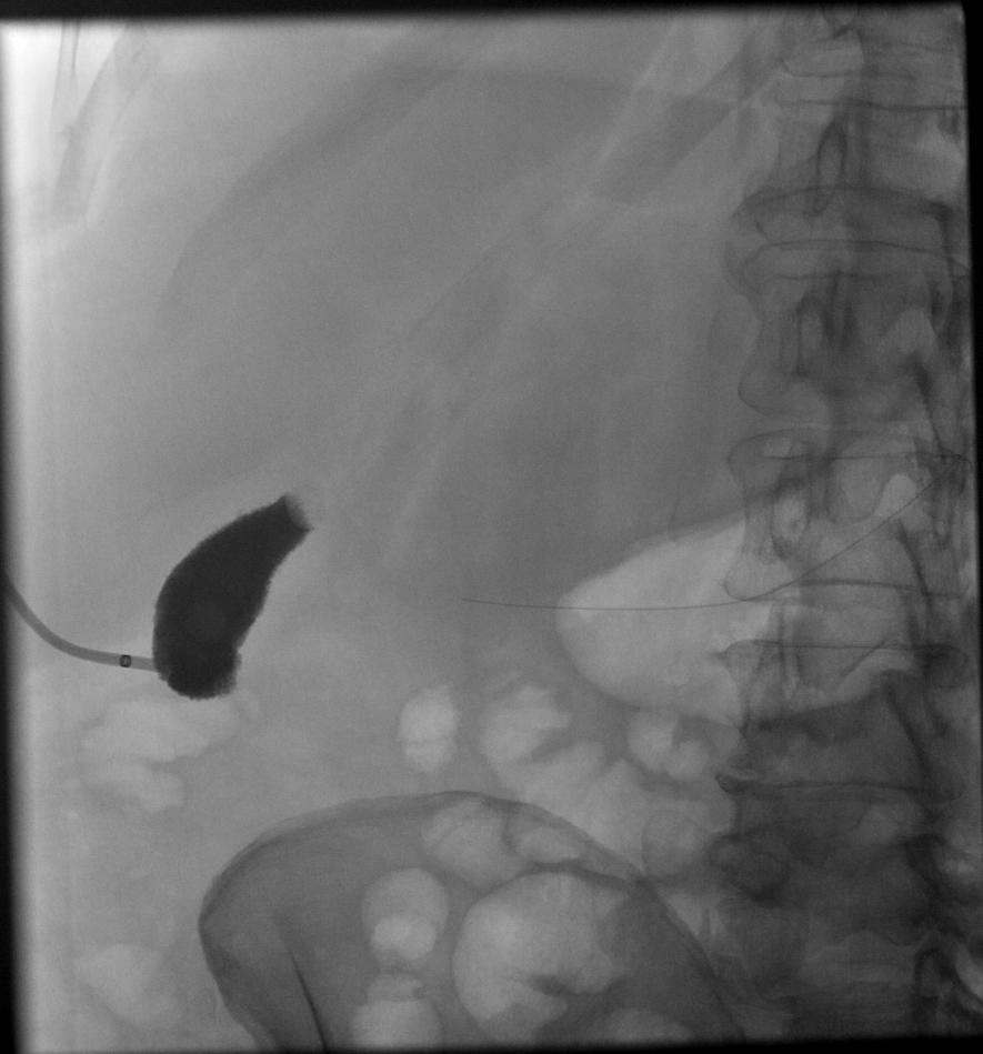
The early views are often the best before you flood the system with contrast so make sure you magnify cone in and obtain good quality images early.
Early image showing multiple filling defects in keeping with gallstones. Note the appearance of a cholecystectomy pigtail catheter.
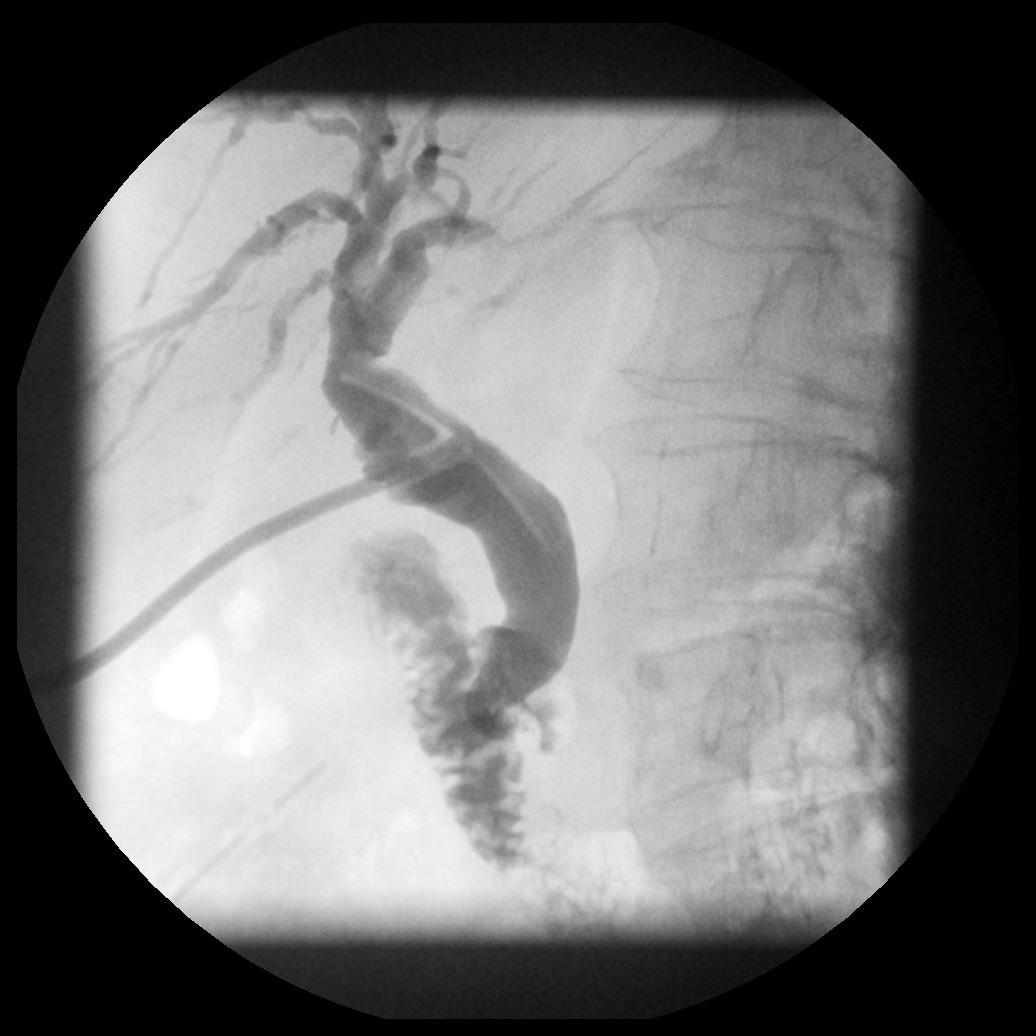
The CBD and left sided ducts often lie over the spine. Oblique imaging e.g LAO is often useful to move the CBD to the right, off the spine.
Once your radiography has been modified continue to inject taking exposures at relevant intervals to demonstrate necessary anatomy and pathology. The most common indications require information regarding correct drain position, leak and patency of the CBD
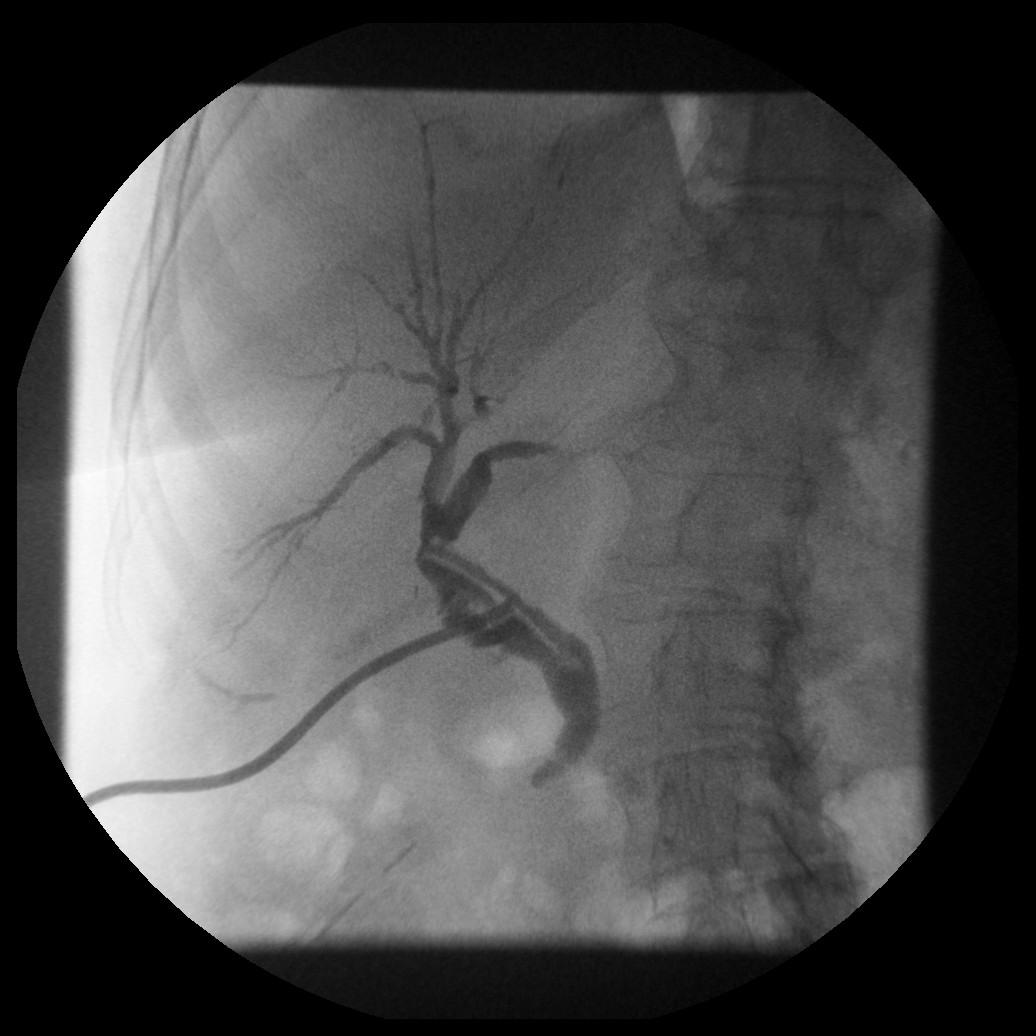
Magnified view showing a correctly positioned T Tube within the CBD. There is no leak. Normal reflux is seen into non dilated intrahepatic ducts. The distal CBD is seen but duodenum not yet visualised.
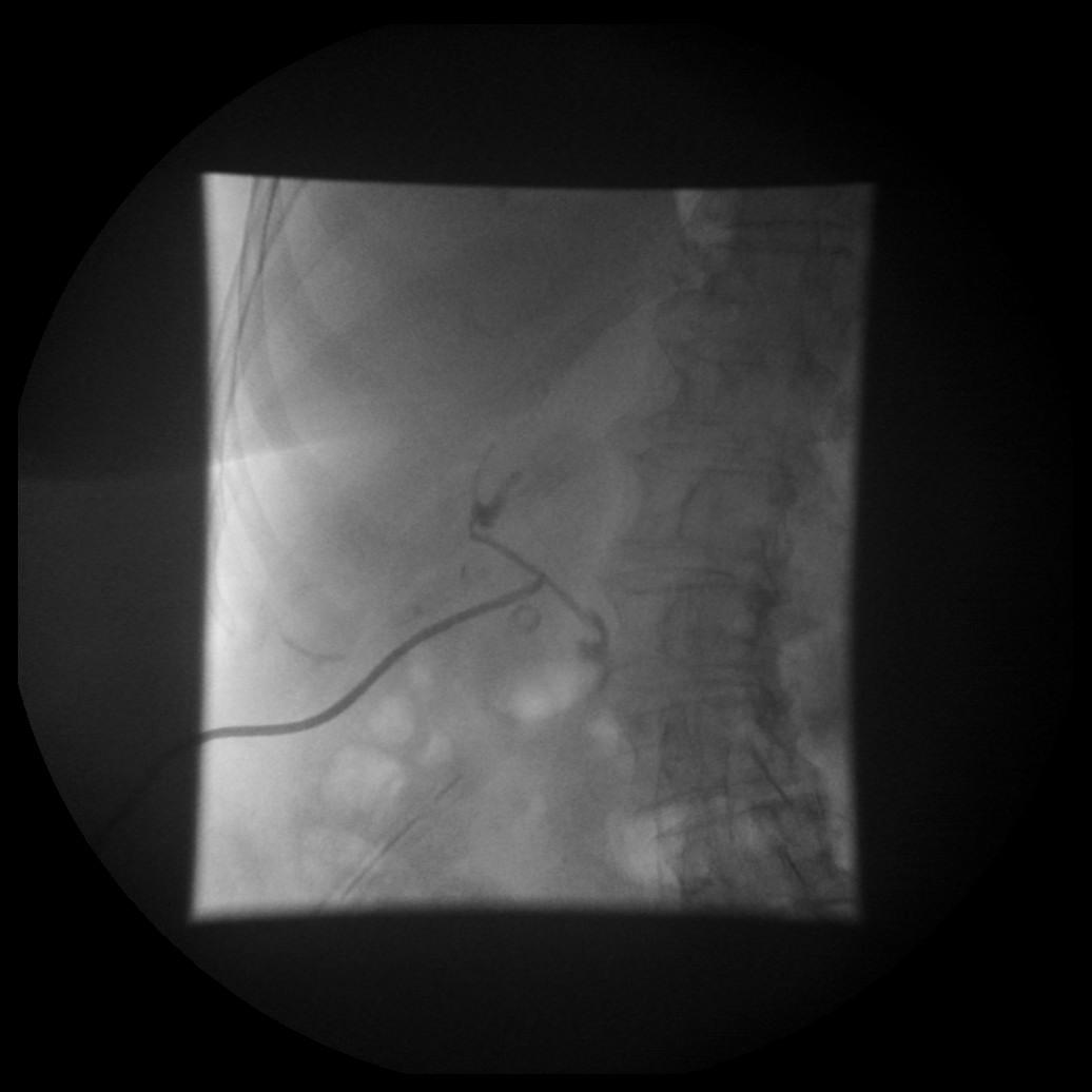
This subsequent image now demonstrates normal flow through the ampulla into the duodenum with no distal obstruction or filling duct. Note the image has been further magnified and centred on the ampulla.
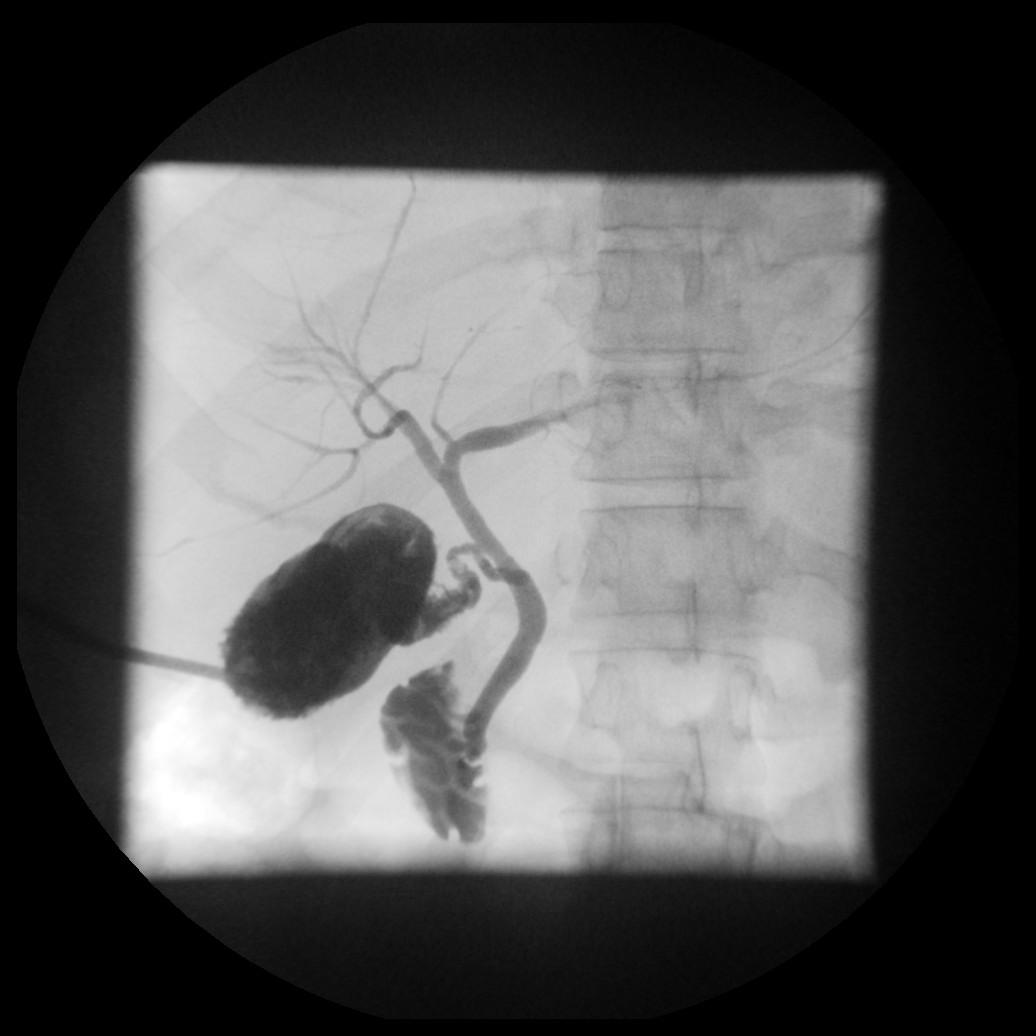
In most instances if the tube is the correct position, is not blocked and the CBD drains into the duodenum with no distal obstruction, the study is finished.
Once complete aspirate the contrast and flush with saline. Keep imaging at this point as diluting the contrast can sometimes show previously unseen calculi. Repeat a post drainage film.
Reconnect or clamp the tube as required.
Assessment
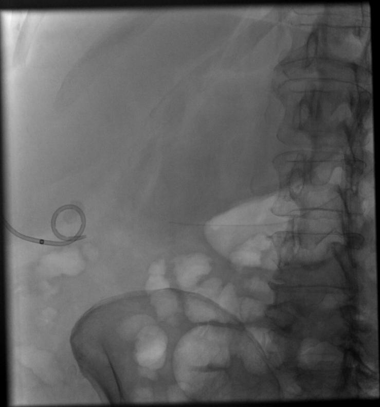
AP control image of a cholecystectomy tube with early contrast injection showing the radiopaque marker lies outside the gallbladder. This is vital to recognise and document, as there is impending displacement of the drain.
Hints and Tips
Ensure you do not inject air, it can mimic calculi. If air is introduced you can try two things, flush air through with saline and repeat cholangiogram with more contrast or try to aspirate the air and repeat the cholangiogram.
If you are unsure if there is a calculus or air, then try flushing or aspirating as above, air will either disappear or move on, a stone will usually remain.
Remember early images. Once the contrast reaches the duodenum, it cannot be aspirated. This contrast can reflux back into the duodenal cap or stomach and obscure detail.
Oblique views are sometimes required e.g. LAO to move the CBD off the spine.
Craniocaudal angulation can also be used to improve images.
On later images the contrast can become quite dense and so small calculi can be hidden. Therefore saline can be used to dilute and flush the contrast further through the biliary tree. This will still provide good quality images. You can always use more contrast afterwards.
Multiple small gallstones become apparent once contrast has been diluted and aspirated.